
FIGURE 5.
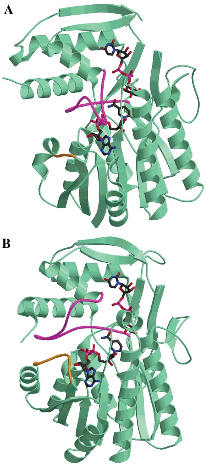
(A) Crystal structure of the E. coli ArnA decarboxylase domain with substrates modeled in the active site. (B) Crystal structure of E. coli UDP-galactose 4-epimerase with its substrates bound in the active site (PDB ID: 1A9Y). The two loops highlighted in magenta and gold in both proteins reveal structural differences likely to be important in substrate binding.