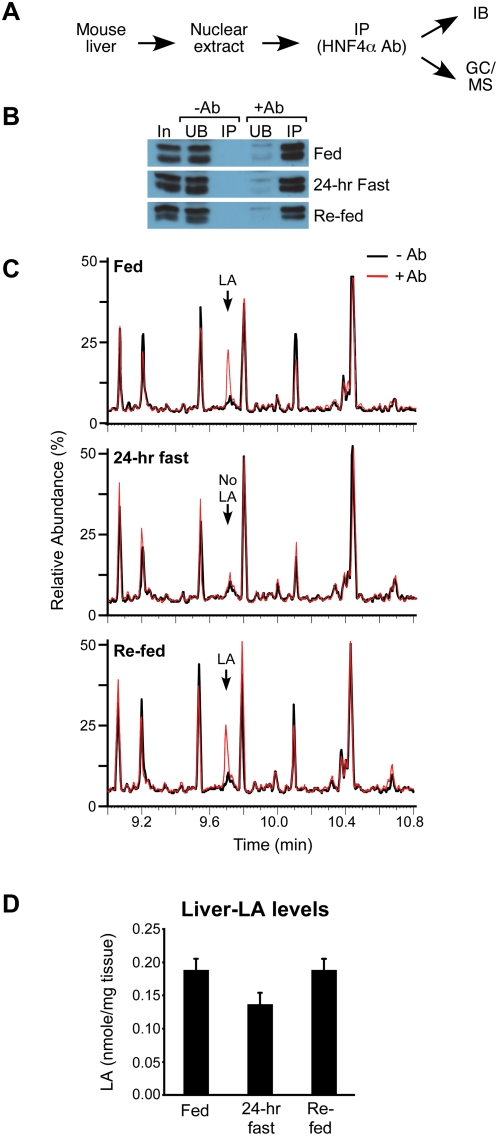
Figure 3. Native hepatic HNF4α binds endogenous LA in fed but not fasted mice.
(A) Design: As in Figure 1A except with livers from male C57BL/6 mice fed a standard diet (Fed), fasted for 24 hr (24-hr Fast) or fasted for 24 hr and re-fed for 24 hr (Re-fed). (B) HNF4α from hepatic nuclei was IP'd in the absence (−Ab) or presence (+Ab) of the HNF4α-specific antibody and quantified by IB. HNF4α resolves as a doublet in this gel system. In, 2% of total input; UB, 2% of unbound material; IP, 2% of IP'd material. (C) GC/MS chromatograms (9 to 11 min) comparing compounds extracted from IP'd material from fed, fasted and re-fed animals with (red) or without (black) HNF4α Ab. Arrow, LA peak. (D) Quantification of the amount of LA in the liver of fed, 24-hr fasted and re-fed mice using GC/MS. Shown are average amounts of LA (nmole/mg liver) from 6 mice +/− SEM per group. Statistics: two-tailed t-test: fed vs. fasted, p = 0.057; fasted vs. re-fed, p = 0.051; fed vs. re-fed, ns; ANOVA p = 0.0641.

