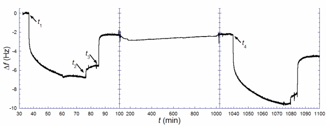
Figure 3.
Changes in f upon addition of anti-biotin (t1 = 37 min) to an SLB from vesicles with biotin-lipids selectively incorporated in the outer leaflet. The exposure was followed by rinsing (t2 = 77 min) and addition of free biotin (t3 = 86 min) to promote removal of bound anti-biotin. Also shown is a second addition of anti-biotin (t4 = 1039 min) followed by rinsing (t = 1079 min) and addition of free biotin (t = 1085 min).

