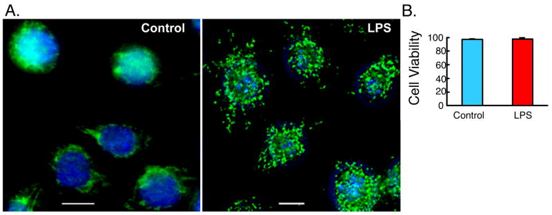
Figure 1. LPS induces formation of autophagosomes.
(A) RAW264.7 cells were incubated in the absence or presence of LPS for 16 h, fixed, stained with DAPI to visualize the nuclei (blue), and immunolabled with anti- LC3 antibody followed by Alexa Fluor 488-conjugated goat anti-rabbit IgG (green). Representative images are shown. (B) Cell viability analysis of RAW264.7 cells, stably expressing LC3-GFP, following incubation in the presence or absence of LPS (100 ng/ml) for 16 h. Cell viability was determined using Cell Viability Analyzer (Vi-Cell, BECKMAN COULTER) based on trypan blue reagent (Mean±SEM, n=4).

