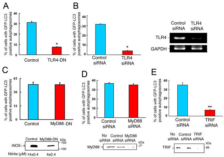
Figure 3. TRIF-dependent TLR-4 signaling is required for LPS-induced autophagy.
Quantitation analysis of the percentage of cells with GFP-LC3-positive autophagosomes in RAW264.7 cells, stably expressing GFP-LC3, after incubation in the presence of LPS (100 ng/ml) for 16 h. In (A), cells were transfected with vector only or with a plasmid expressing TLR4-dominant negative mutant. In (B, D and E), cells were transfected with control siRNA or siRNA specific for TLR4, MyD88 or TRIF, respectively. All transfections were done for 32 h, prior to LPS treatment. In (C), cells stably expressed both GFP-LC3 and MyD88 dominant negative mutant. Right panel in (B)- results of RT-PCR confirming deletion of TLR4 by siRNA. Lower panel in (C)- evaluation of iNOS protein expression by Western analysis and iNOS activity by measuring nitrite in culture media. Lower panel in (D)- Western analysis of MyD88 in cell lysates. Lower panel in (E)- Western analysis of TRIF in cell lysates. Data represent means±SEM of three independent experiments. * and ** denote p < 0.05 and p < 0.001, respectively, when compared to control condition.

