
Figure 7. p38 MAPK inhibition blocks LPS-induced co-localization of Mycobacterium tuberculosis with autophagosomes.
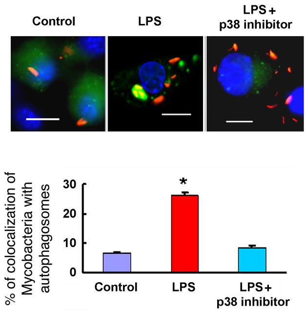
RAW264.7 cells stably expressing GFP-LC3 were infected with Mycobacterium tuberculosis expressing red fluorescent protein (RFP) 1 h prior to a 30 min incubation with p38 MAPK inhibitor or vehicle (DMSO) followed by further incubation for 16 h in the presence or absence of LPS. Upper panel: representative fluorescence images. Lower panel: Quantitation of percentage of colocalization of Mycobacterium tuberculosis with GFP-LC3-positive autophagosomes GFP-LC3-positive autophagosomes. Graph represents the quantitation of 100 internalized mycobacteria per experimental condition. Data denote means ± SEM from two independent experiments. Scale bar, 10 μm. *denotes p< 0.05, when compared to control condition.