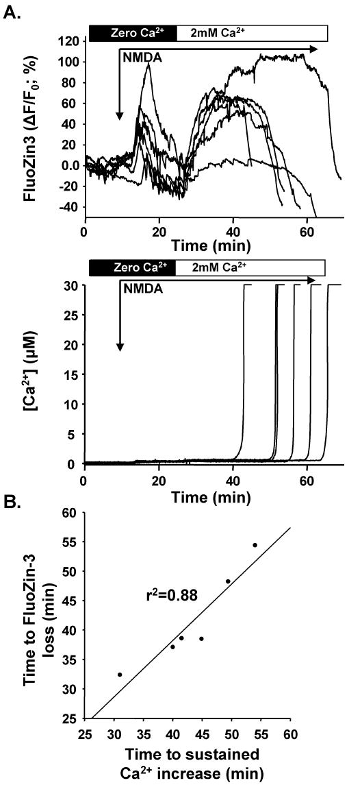
Figure 6. Effects of extracellular Ca2+ removal.
A: Near-simultaneous FluoZin-3 (top) and Fura-6F (bottom) records from co-loaded neurons. Extracellular Ca2+ was washed out for 10 min before exposure to NMDA in the nominally Ca2+-free solution. Subsequently 2mM Ca2+ was introduced 15 min into the NMDA exposure. In the nominal absence of extracellular Ca2+, initial FluoZin-3 increases were reduced but not abolished (compare with Figure 4). Reintroduction of Ca2+ produced prompt and sustained FluoZin-3 fluorescence increases. The lower panel of A shows that Ca2+ removal virtually abolished Fura-6F responses, until sustained increases were observed after reintroduction of Ca2+. B: As in control conditions, late FluoZin-3 loss was coincident with arrival of sustained Ca2+ increases in somata. The relationship between the time at which 50% FluoZin-3 fluorescence intensity (compared with plateau levels prior to arrival of Ca2+ overload) is lost is plotted against the time of half-maximal Ca2+ increase as the sustained Ca2+ response invaded somata for the 6 neurons shown in A.

