
Figure 4.
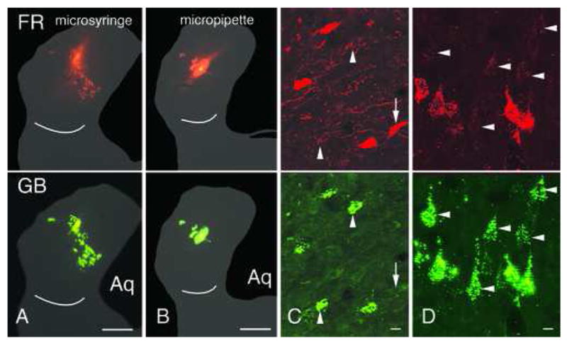
Photomicrographs of injection sites and retrogradely labeled cells after co-injections of FluoroRuby (FR) and green beads (GB). A, B. Co-injections made with a microsyringe (A) or a micropipette (B). C, D. Labeled cells in the right inferior colliculus (C) or left temporal cortex (D). Cells are double- or single-labeled. Arrows: cells that contain only FR; arrowheads: cells that contain only GB. Scale bars: 1 mm (A, B); 10 μm (C, D). Aq –cerebral aqueduct. Cases GP433 (A); GP436 (B–D).