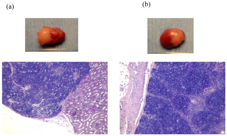
Figure 6.
Histology and gross appearance of thymic grafts obtained at 20 weeks post-implantation from a NOD/SCID mouse transplanted with both human and swine thymus grafts under opposite kidney capsules. Both human (a) and swine (b) thymic grafts showed normal thymic architecture with readily visible cortical and medullary structure (H-E staining).

