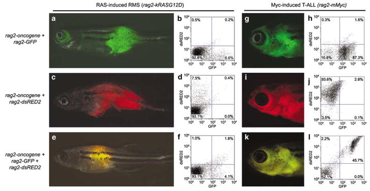
Figure 1.
Co-injection of multiple transgenes into one-cell stage zebrafish embryos leads to co-expression in tumors. Rhabdomyosarcoma (RMS) was induced by injection of rag2-kRASG12D (a–f), whereas T-cell acute lymphoblastic leukemia (T-ALL) was induced by injection of rag2-mMyc (g–l). Oncogene-containing transgenes were co-injected with rag2-GFP (a, b, g and h), rag2-dsRED2 (c, d, i and j) or both (e, f, k and l). Fluorescent microscopic images show expression of GFP (a and g), dsRED2 (c and i) or both (e and k). FACS analysis confirms expression patterns of tumor cells from animals shown in the corresponding fluorescent images (b, d, f, h, j and l). The rag2-GFP (Jessen et al., 2001), rag2-mouse cMyc (Langenau et al., 2003), rag2-dsRED2 and rag2-human kRASG12D (Langenau et al., 2007) constructs have been described previously. DNA constructs were linearized with either XhoI or Not1, phenol:chloroform extracted and ethanol precipitated as described previously (Langenau et al., 2003). For rag2-oncogene, rag2-dsRED2 co-injections (a, b, g and h), the oncogene containing construct was linearized with XhoI and the second linearized with NotI. For rag2-oncogene co-injections with rag2-GFP (c, d, i and j), both constructs were digested with the same enzyme, either NotI or XhoI. DNA was quantified by both spectrophotometric analysis and gel quantification. DNA was diluted in 0.5XTE + 100 mM KCl to 100 ng μl−1, with co-injection of two transgenes having 50 ng of each construct per microliter or three transgenes having 33.3 ng of each construct per microliter. FACS, fluorescence activated cell sorting; GFP, green fluorescent protein.

