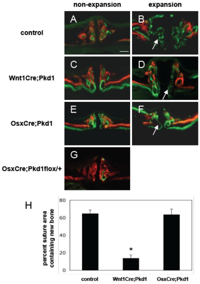
Figure 1.

Deficient new bone formation in response to expansive force in Wnt1Cre;Pkd1 mice. All mice were injected with alizarin complexone (red) and calcein (green) 15 days and 1 day before euthanasia, respectively. Frontal sections of maxillae of control (A and B), Wnt1Cre;Pkd1 (C and D), and OsxCre;Pkd1 (E and F) mice with (B, D and F) or without (A, C and E) application of the opening loops. (G) The thickness of midpalatal suture and palatal bones of OsxCre;Pkd1flox/+ mice is comparable to that of controls. (H) Comparative analysis of percent suture area containing new bone in control group, Wnt1Cre;Pkd1 group and OsxCre;Pkd1 group at day 14 (*p<0.001). (B, D and F) Arrows point to new bone formed in the expanded suture area during the experiment period of 2 weeks. Scale bar (A): 100μm.
