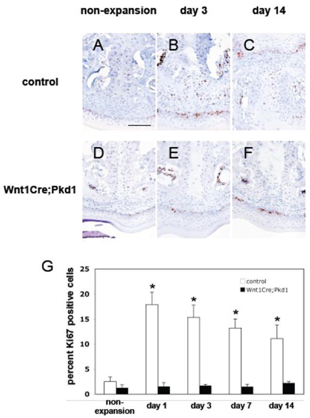
Figure 3.

Deficient periosteal cell proliferation in Wnt1Cre;Pkd1 mice in response to expansive force. Ki67 immunohistochemical staining of frontal sections of midpalatal sutures of control (A-C) and Wnt1Cre;Pkd1 (D-F) animals without expansion (A and D) and with expansion at days 3 (B and E) and 14 (C and F). (G) Comparative analysis of Ki67 expression in control groups and Wnt1Cre;Pkd1 groups without expansion and with expansion at days 1, 3, 7 and 14 (*p<0.05). (B) Ki67-positive cells (brown) are located in the periosteal region. (C) Ki67-positive cell are distributed within the expanded suture. (F) Ki67-positive cells are located in the periosteal region. Scale bar (A): 100μm.
