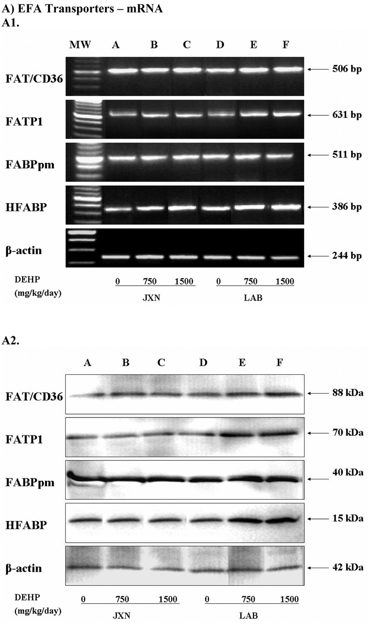
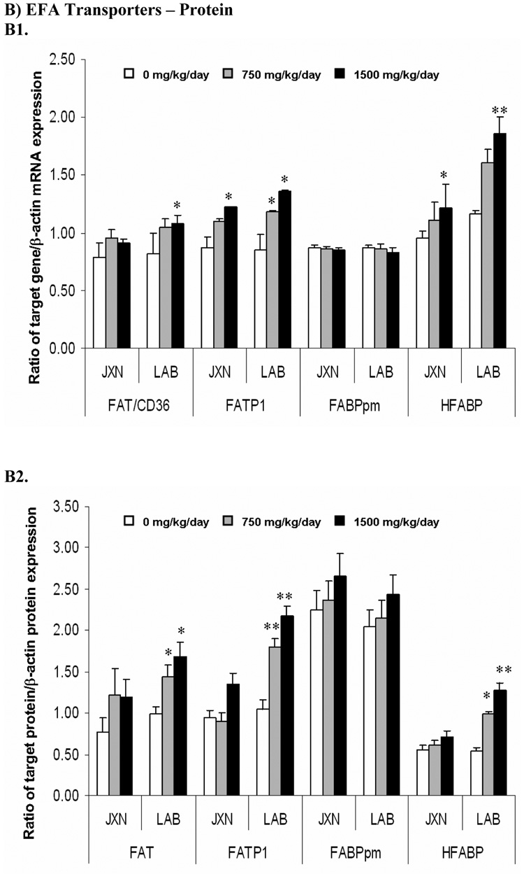
Figure 2. Expression of fatty acids transporters mRNA (Panel A) and protein (Panel B) in rat placenta upon maternal exposure to DEHP.
Dams were dosed with DEHP (750 mg/kg/day or 1500 mg/kg/day) or vehicle from GD 0 through GD 19. Total RNA and whole cell protein were isolated at GD 20 and expressions of FAT/CD36, FATP1, FABPpm and HFABP mRNA and protein were analyzed using semi-quantitative RT-PCR and Western blot, respectively. A1 and B1, representative RT-PCR gel electrophoresis and Western blot image. Lane A-C: JXN, junctional zone; Lane D-F: LAB, labyrinthine zone. A2 and B2, densitometry analysis of the RTPCR gel and Western blot image. Data shown were relative arbitrary values after normalized with β-actin (means ± SD, n =3 or 4). *, p<0.05; **, p<0.01; ***, p<0.001.


