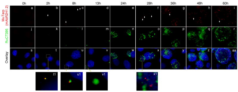
Fig. 3. Time course expression of Tarp.
HeLa cells infected with C. trachomatis serovar D organisms were processed at various time points after infection as indicated on top of the figure. The infected cell monolayers were immunostained with a mouse anti-Tarp mAb plus a goat anti-mouse IgG conjugated with Cy3 (red) and a rabbit anti-chlamydial organism antibody plus a goat anti-rabbit IgG conjugated with Cy2 (green) and a DNA dye (blue). Please note that Tarp was detected during the first 8 hours after infection (panels t1 & u1) and disappeared (panels v1 & w) until 28 hours after infection (×1). As incubation continued, more Tarp-positive granules were detected (panels g to i).

