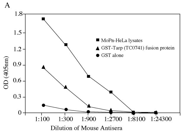
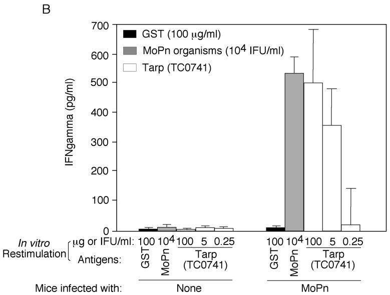
Fig. 5. Induction of Tarp-specific immune responses by MoPn intravaginal infection.
Mice were intravaginally infected with or without MoPn organisms (1 × 10⁴ IFU) as described in the method and material section and 30 days after infection, both sera were collected for antibody measurements (A) and spleen cells for in vitro stimulation assay (B). For antibody measurement, the pooled serum samples from each group were serially diluted as shown along the X-axis and assayed against MoPn-HeLa lysates (filled square), GST-Tarp fusion protein (filled triangle) or GST alone (filled circle) in ELISA. The results were expressed as OD values as shown along the Y-axis. Please note that the mouse antisera positively reacted with both MoPn antigens and Tarp but not GST alone. For monitoring cellular response, the spleen lymphocytes harvested from the mice were re-stimulated in vitro with the following antigens as listed along the X-axis: UV-inactivated MoPn EB organisms at 1 × 10⁴ IFUs/well, GST-Tarp fusion protein at the concentrations of 100, 5 & 0.25ug/ml and GST alone at 100ug/ml. Three days after the stimulation, the culture supernatants were collected for IFNg detection in an ELISA and the results were expressed as pg/ml as listed along the Y-axis.


