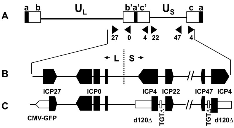
Figure 1. Organization of the HSV-1 genome.
A.Diagram of the structure of the HSV genome. The unique sequences are represented as a line, and the repeated sequences are represented as boxes. a= terminal repeats, b, b′ = L component inverted repeats, c, c′= S component inverted repeats; B. Expanded map of the right end of the genome showing the ORFs of the IE genes; C. Expanded map of the right end of the d106 genome showing the deletions (open boxes) of ICP4 and ICP22/47 gene promoters, and the CMV-GFP cassette (open arrow) insertion in the ICP27 gene.

