
Fig. 4. ChIP analysis of PAX2 binding to the hBD1 promoter, in vivo.
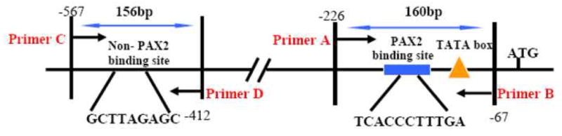

Chromatin immunoprecipitation- assays were performed to examine PAX2 binding activity in situ using DU145 and PC3 cells. A, PCR amplification of precipitated DNA was performed with primers flanking the PAX2 binding motif (A and B) or a region null for the PAX2 recognition sequence (C and D). B, Whole cell lysates from formaldehyde cross-linked DU145 or PC3 cells were incubated with antibodies and DNA-protein-antibody complexes were recovered by incubation with Protein A sepharose beads. The presence of hBD1-DNA in immunoprecipitated complexes was detected by PCR using primer sets A and B. PCR products were analyzed on a 2% agarose gel, which was stained with ethidium bromide. Precipitation without antibody (lane 1) or with normal IgG (lane 2) resulted in no amplification product. However, amplification of DNA precipitated with anti-PAX2 antibody generated 160 bp products in DU145 cells (lane3) and PC3 cells (lanes 3–4).