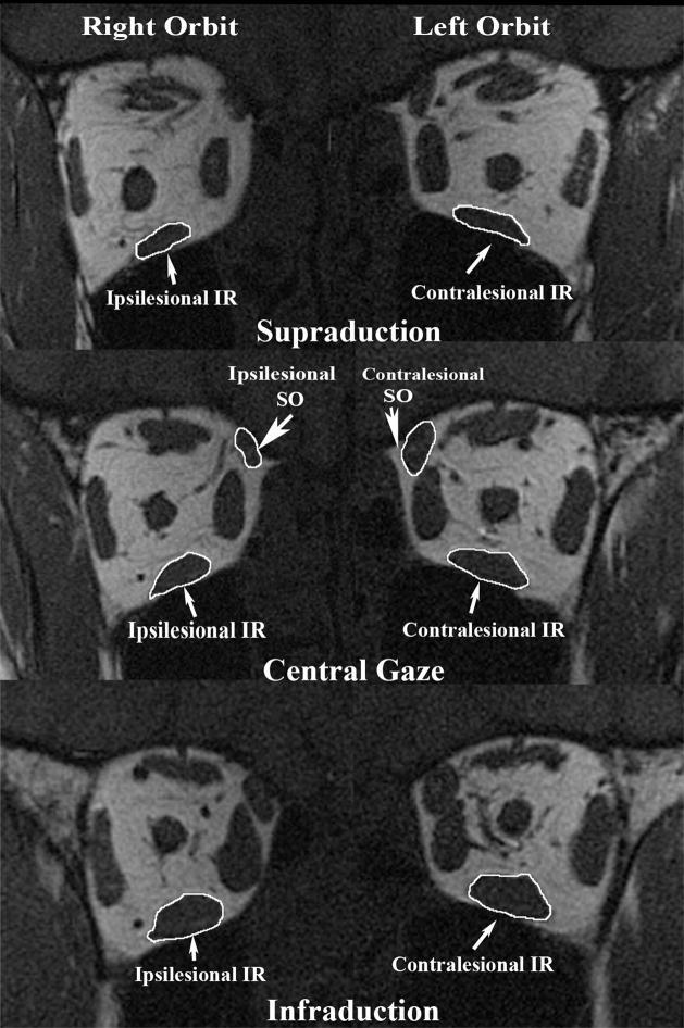
Figure 1.
Quasi-coronal MRI 8 mm posterior to the globe-optic nerve junction (ON) in supraduction, central gaze, and infraduction in subject 5 with congenital right SO palsy. Note smaller cross-section of right than left SO and less contractile thickening of right SO from supraduction to infraduction. Also note smaller cross-section of right than left IR and less contractile thickening of right IR from supraduction to infraduction. IR = inferior rectus muscle; SO = superior oblique muscle.

