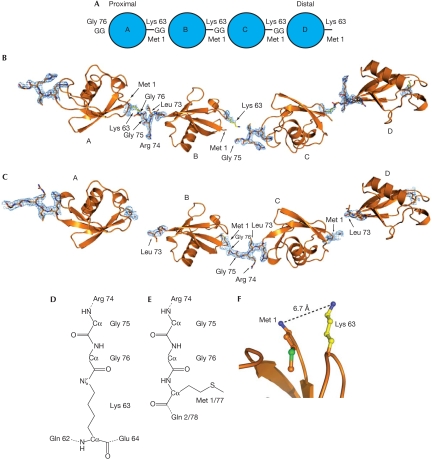
Figure 1.
Structure of Lys 63 and linear ubiquitin chains. (A) Nomenclature for polyubiquitin chains. The proximal molecule is linked through its carboxy terminus to a substrate lysine residue, or has a free carboxy-terminal diGly (GG) motif in unattached chains. (B,C) Four equivalent ubiquitin molecules, corresponding to two adjacent asymmetric units within the crystal lattice, are shown in cartoon representation. 2∣Fo∣−∣Fc∣ electron density at 1σ is drawn for the linkage residues between molecules A–B and C–D for Lys 63-linked diubiquitin, and for B–C in linear diubiquitin. (D) Chemical representation of the Lys 63 linkage. Other isopeptide linkages (for example, Lys 48 linkages) differ only in the type of neighbouring residues. (E) Representation of the peptide linkage in a linear ubiquitin chain between Gly 76 and Met 1 of the second molecule. (F) Close spatial location of Lys 63 and Met 1 (distance of 6.7 Å) allow similar conformation of linear and Lys 63-linked chains. Ub, ubiquitin.

