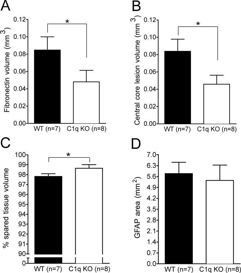
Figure 6.
Unbiased stereological quantification of pathology 28 d post-SCI. A, C1q KO mice showed a significant reduction in fibronectin volume compared with WT mice (Student's t test, *p < 0.05). B, This finding was corroborated by the significant reduction in the central core lesion volume observed in C1q KO mice compared with WT mice (Student's t test, *p < 0.05). C, Additionally, C1q KO mice showed a significant increase in spared tissue volume compared with WT mice (Student's t test, *p <0.05). D, However, the GFAP area was not significantly different between the groups (Student's t test, p > 0.05). Error bars represent group means ± SEM.

