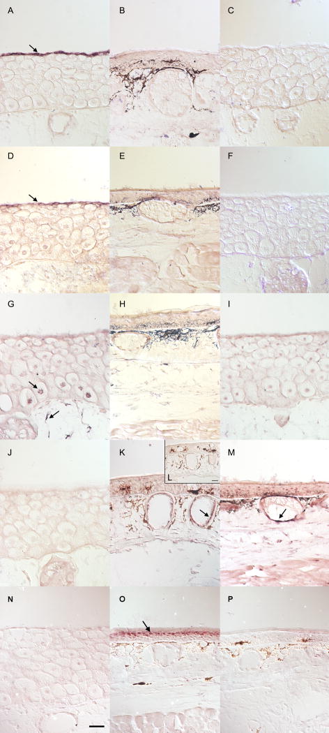
Fig. 8.
In situ hybridizations of metamorphosing head skin. Control-sense probes were run on slides collected from the same animal as the positive signal. Black deposits in panels B, E, H, K, L, O, and M, and P are melanin deposits and not positive signal. Anti-sense Day 0 samples can be found in panels A, D, G, J, and N. An Anti-sense Day 12 sample can be found in panel K. Anti-sense Day 28 samples can be found in panels B, E, H, M and O. Control-sense Day 0 samples can be found in panels C, F, and I. Control sense Day 12 and Day 28 samples can be found in panels L and P, respectively.. UMOD (A–C) and KRT6A (D–F) are exclusively expressed in the apical cells of larval skin (A and D arrows). CALM2 (G–I), is expressed in basal Leydig cells and highly expressed in dermal fibroblasts (panel G, arrows). MMP1 (J–M) is highly expressed in dermal glands on Day 12 (K, arrow) and Day 28 (M, arrow). KRT14 (N–P) is highly expressed in the granular layer of Day 28 epidermis (O, arrow). The scale bar is 50 μm.

