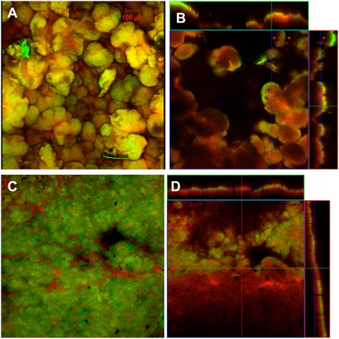
Figure 1. Confocal scanning laser microscopy images of Geobacter sulfurreducens grown with different electron acceptors.
Confocal scanning laser microscopy images of current harvesting and fumarate control biofilms of wild type G. sulfurreducens. Metabolically active (green) and inactive (red) cells where differentiated with a LIVE/DEAD kit based on the permeability of the cell membrane. A. 3-D projection, top view, fumarate control biofilm; B. slices through biofilm parallel to electrode large panel and perpendicular to electrode top and side panel, fumarate control biofilm; C. 3-D projection, top view, current harvesting biofilm; D. slices through biofilm parallel to electrode large panel and perpendicular to electrode top and side panel, current harvesting biofilm.

