
Figure 5.
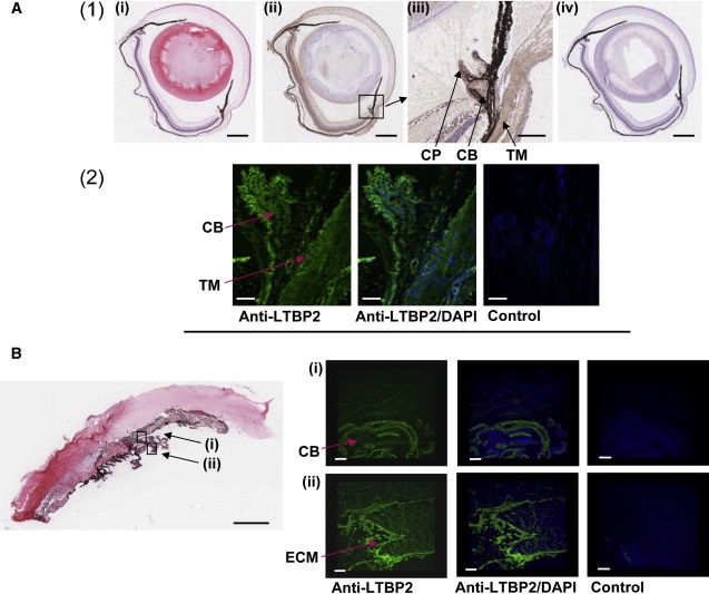
Localization of LTBP2 in the Adult Eye
(A) (1) Formalin-fixed sections of adult mouse eyes stained with hematoxylin and eosin (i), anti-LTBP2 (ii), or the Rabbit Envision Detection system (iv). In (ii), LTBP2 immunoreactivity was observed as brown staining in several structures of the eye including the sclera, retina, cornea, trabecular meshwork, and the ciliary body. The black intense pigment that extends from the retinal pigment epithelium to the iris is melanin (in (i), (ii), and (iv)). Scale bars represent 500 μm. The area surrounding the ciliary body in (ii) has been magnified and is shown in (iii). Note LTBP2 immunoreactivity particularly at the trabecular meshwork (TM), the ciliary body (CB), and the ciliary process (CP). Scale bar represents 100 μm. (2) Cryopreserved sections of adult mouse eyes stained either with anti-LTBP2 or a rabbit IgG isotype negative control. LTBP2 immunoreactivity depicted as green fluorescence was present in the trabecular meshwork (TM), the ciliary body (CB), and particularly the ciliary process. Scale bars represent 50 μm.
(B) Cow eye histological section showing the cornea, iris, and ciliary body stained with hematoxylin and eosin. Scale bar represents 1500 μm. The lens dislocated during the sectioning and the adjacent alignment of the iris with the cornea in the panel is an artifact of tissue preparation. Cryosections stained with anti-LTBP2 or rabbit IgG negative control are shown. Note that the green fluorescence corresponding to LTBP2 immunoreactivity is abundant in the ciliary body (CB) (i) and particularly intense in the extracellular matrix structures (ECM) at the ciliary process (ii). Scale bars represent 150 μm.