
Figure 1.
Histochemical and Ultrastructural Analysis of Probands' Skeletal Muscle
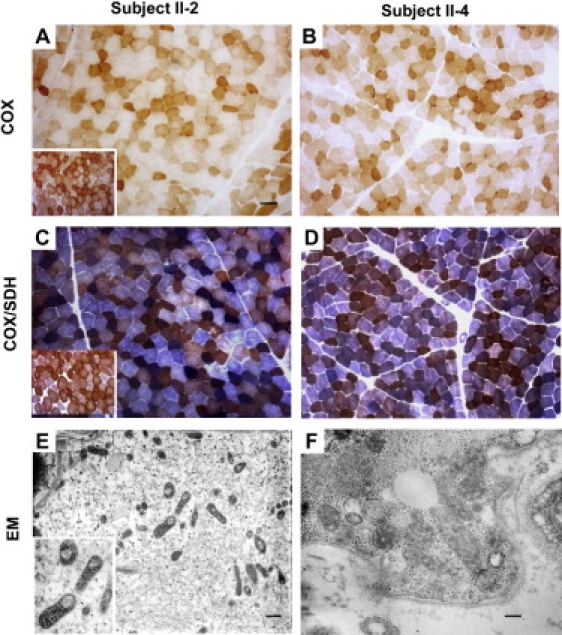
(A–D) COX histochemistry reveals the presence of several COX-deficient fibers in probands II-2 and II-4 (A and B). Double staining for COX/SDH. Scattered fibers with increased SDH staining, indicative of mitochondrial proliferation, are present in patient II-2. A COX deficiency is confirmed in both patient II-2 and patient II-4. (C and D) Insets within panels (A) and (C) represent the normal controls.
(E and F) Electron microscopy shows several mitochondria with thickened cristae; many mitochondria are also present with large vacuolization in (E).
Scale bars represent 25 μm (A–D), 700 nm (E), or 250 nm (F).