
Figure 3.
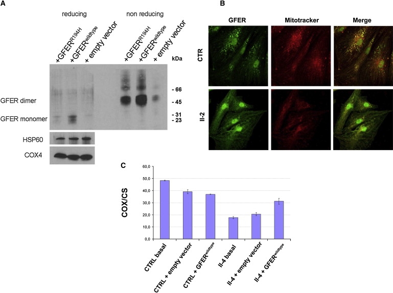
Western Blot Analysis of Overexpressed GFER, Immunocytochemical Analysis of Endogenous GFER in Patient Myoblasts, and Functional Rescue in Patient Fibroblasts
(A) HEK293 cells were stably transfected with vector overexpressing GFERwild-type and GFERR194H cDNA. Immunoblot analysis showed a reduction of GFERR194H in mitochondrial fractions under both reducing (DTT 15 mM) and nonreducing conditions. COX4 and HSP60 were used for normalization.
(B) Antibodies against human GFER showed specific GFER colocalization with MitoTracker and DAPI in control myoblasts, whereas immunofluorescence was almost absent in the cytoplasm. Instead, primary myoblasts from patient II-2 showed a reduced punctuate mitochondrial fluorescence compared to control cells with more diffuse cytoplasmic staining. Scale bar represents 40 μm.
(C) Biochemical functional rescue in proband II-4 fibroblasts that were transfected with GFERwild-type cDNA. The analysis of COX activity (normalized to citrate synthase [CS]) shows a restoration of mitochondrial cytochrome c oxidation (complex IV). Results are presented as mean of triplicate determinations ± SD.