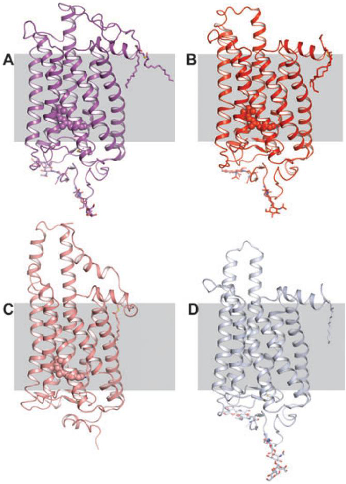
Figure 2.
Extension of the C-III loop of rhodopsins when placed in the context of a hypothetical membrane. (A) Rhodopsin structure solved in the tetragonal space group P41 (PDB ID:1U19). (B) Rhodopsin solved in trigonal space group P31 and re-refined in the higher symmetry space group P64 (PDB IDs:1GZM and 3C9L respectively). (C) Squid rhodopsin (PDB ID:2Z73). (D) Opsin solved in the trigonal space group, H3 (PDB ID:3CAP). A large protrusion of the C-III loop is evident in B, C and D and this region has higher temperature factors than the surrounding residues and is disordered in the photoactivated structure (PDB ID:2I37). It is rigidified in the opsin structure by interaction with C-I on a symmetry-related molecule. It is likely that this region must move or become less ordered upon activation in order for Gt to interact with and become activated by the receptor.

