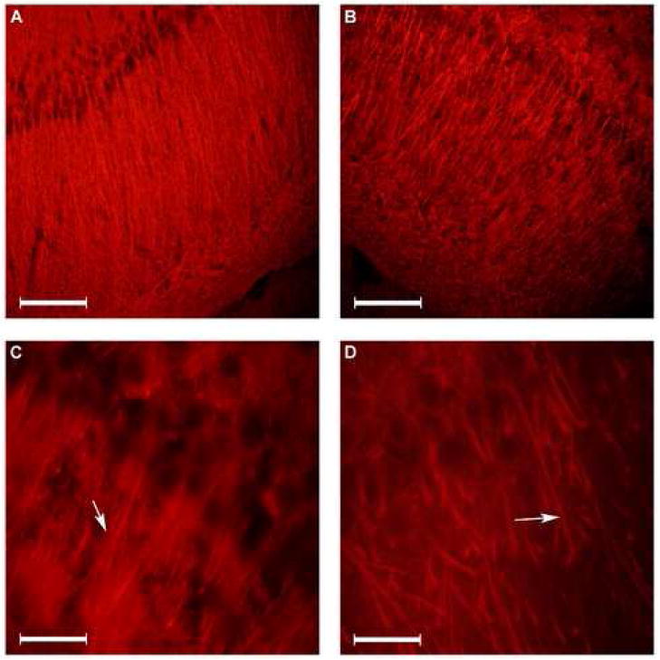
Figure 1.
Microtubule Associated Protein (MAP) -2 immunofluorescence staining of hippocampal CA1 subarea. The dendritic morphology in the hippocampal CA1 subarea in normoxic (panels A and C) and chronic hypoxic (panels B and D) rats using MAP-2 immunofluorescence is shown. Arrows point to the origin of initial branching. (20μm coronal sections of the dorsal hippocampus. Bar in panels A and B = 100 μm. Bar in panels C and D = 20 μm).

