Figure 4.

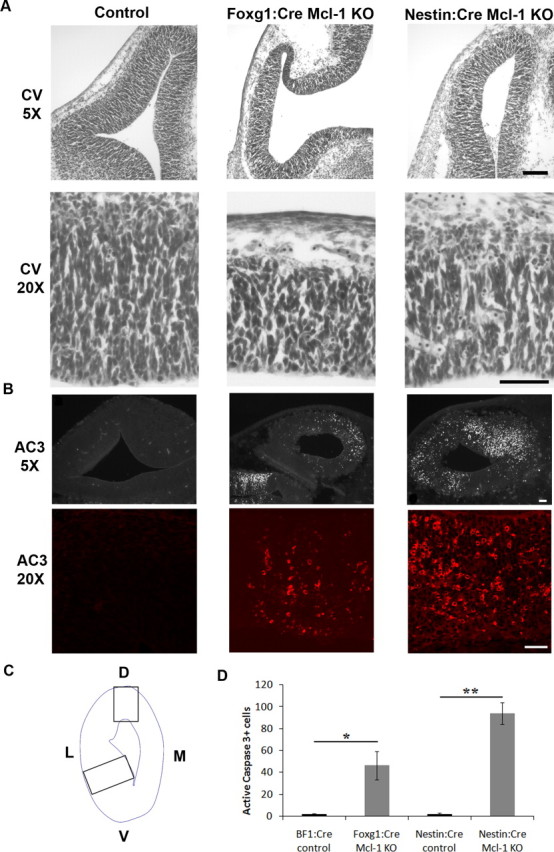
Loss of Mcl-1 in developing neurons results in increased apoptotic activity in neuronal progenitors. A, Cresyl violet-stained coronal sections through the telencephalic hemisphere of E12.5 Foxg1:Cre and Nestin:Cre Mcl-1 mutant embryos and littermate controls showing a significant reduction in the thickness of the developing cortex in the Mcl-1 mutants. The bottom panel represents high-magnification photomicrographs of the developing cortex. B, Coronal sections from E12.5 Foxg1:Cre and Nestin:Cre Mcl-1 mutants and littermate controls were immunostained for AC3, a marker of apoptotic cell death. Numerous active caspase-3-positive cells are dispersed throughout the VZ in Mcl-1 deficient mice. The bottom panels are higher-magnification photomicrographs. C, Schematic representation of areas quantified. D, Quantitative analysis of AC3-positive cells reveals a significant increase in apoptotic cells in mutant animals. L, Lateral; M, medial. Scale bars: A, 100 μm; B, 50 μm. *p < 0.05, **p < 0.01.
