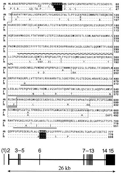
Figure 1.
Amino acid sequence alignment of mouse, rat, and human Sema W and genomic structure of mouse Semaw. (Upper) Residues in the rat and human sequence that are identical to the mouse sequence are represented by dots; gaps in sequence relative to the mouse sequence are represented by dashes. The sema domain (solid outline), Ig-like domain (dashed outline), and the transmembrane domain (single underline) are shown. A region of homology to the vaccinia virus protein A39R is indicated by a wavy underline. A subdomain of plexin/SEX homology is indicated by a double underline. The string of leucines encoded by the CTG repeat is indicated by black shading, and a cyclic nucleotide-dependent phosphorylation site is indicated by dark-gray shading. (Lower) The relative positions and sizes of the Semaw exons in the mouse genomic sequence are shown. Exon 1 has been found thus far only in the rat. Intron lengths over 2,000 bp are estimates calculated by comparing intron-spanning PCR products with size markers on an agarose gel.

