Figure 2.
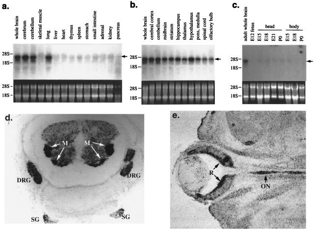
Northern and in situ hybridization analysis of Sema W expression. Northern blots were performed with a rat Sema W probe against a panel of total RNA extracts from various adult rat tissues (a), various adult rat central nervous system tissues (b), and rat embryonic (E) and postnatal (P) tissues at various stages of development (c; given in days post coitus or days after birth, respectively). Below each Northern blot image, confirmation of equivalent RNA amounts by ethidium bromide staining is shown. Locations of 28S and 18S ribosomal RNA are indicated. Sema W bands are indicated with arrows. (d) In situ hybridization with a rat Sema W probe of a transverse section of an embryonic-day-15 rat spinal column. Darkly staining regions are the motor neurons (M), dorsal root ganglia (DRG), and sympathetic ganglia (SG). (e) In situ hybridization with a rat Sema W probe of a coronal section of an embryonic-day-15 rat eye. Staining is seen in the retina (R) and along the optic nerve tract (ON).

