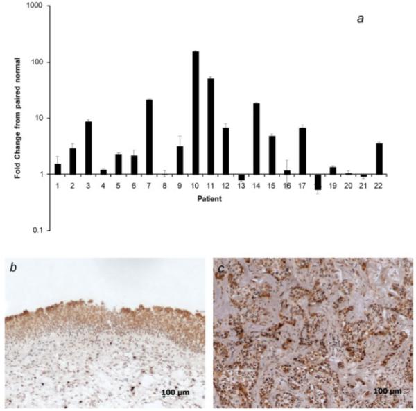
Figure 1.

MUC1 expression in bladder carcinoma. (a) mRNA MUC1 expression was determined in paired normal/cancer samples by realtime RT-PCR. Graph shows fold change in MUC1 expression in tumor sample compared to paired normal tissue sample. Mean ± SD of 3 determinations is shown. The β2-microglobulin gene was used as an internal control. (b and c) Immunohistochemical analysis of section from normal (b) and tumor (c) sample. Sections were stained for MUC1 using anti-MUC1 mAb BCP8. The panels show that MUC1 expression is restricted to the luminal surface of normal urothelium, but is present in the luminal, intermediate and basal layers of the tumor sample. Magnification ×100. Representative images shown (n = 9).
