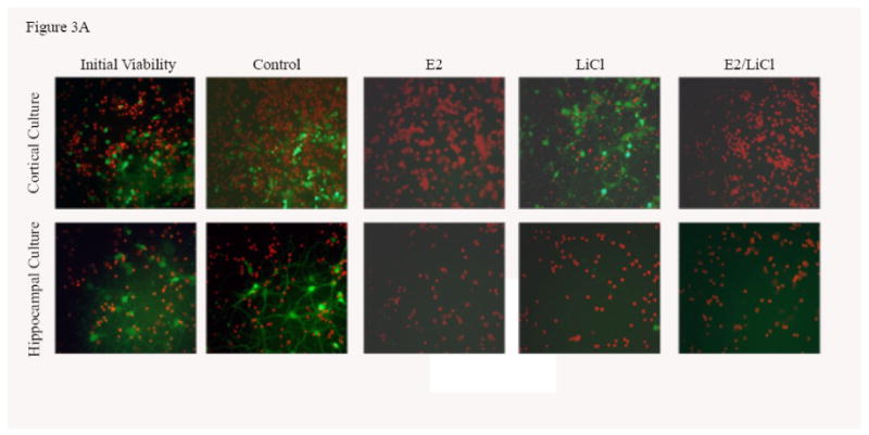
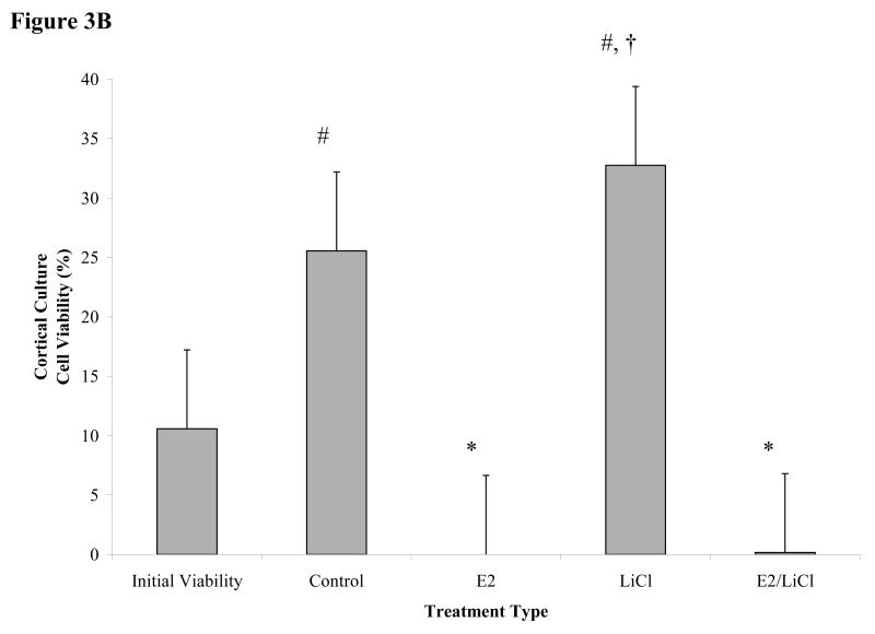
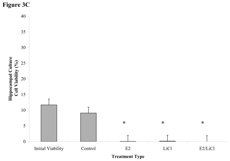
Figures 3 A-C.

Mean viability for glutamate excitotoxicity from 48 h treated cortical and hippocampal cell cultures. Excitotoxicity was induced by incubating primary cultures with 100μM glutamic acid for 1 h at 37°C after pre-treating for 48 h with with 0.04μM E2, 10mM LiCl, combined E2/LiCl, or Control (no treatment). A pre-treatment measurement (Initial viability) was assayed to note any changes caused by the treatment and glutamate excitotoxicity. Images (A) were captured (green labeling, FDA (live cells) and red labeling, PI (dead cells)) and viability (FDA/(FDA+PI)) was assessed using ImageJ Cell Counter for both cortical (B) and hippocampal (C) cultures. *, significantly reduced cell viability compare with Initial Viability and Control (p-value < .05); #, increased cell viability when compared with Initial Viability (p-value < .05); †, increased cell viability when compared with other treatments (p-value < .05).


