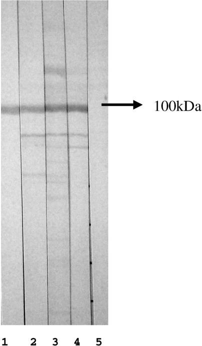
Fig. 3.
Western blot with whole HEp-2 cell extract separated in 10% SDS–PAGE and probed with sera diluted in the ratio 1 : 50. Lane 1: western blot-based affinity-purified antibodies reacting with DNA topo I; lanes 2–4: representative sera depicting the Scl-70 pattern on IIF in HEp-2 cells; lane 5: negative control. Arrow indicates proteins migrating with mobility compatible with 100 kDa.

