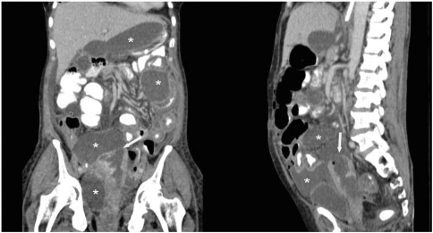
Figure 2.
Contrast-enhanced coronal and sagittal reformatted CT image depicting dilated endometrial cavity with site and size of the breach at uterine fundus (arrow). Multiple pelvic and intra-abdominal collections (*) are seen. The larger pelvic collection is seen in continuation with the ruptured uterus.

