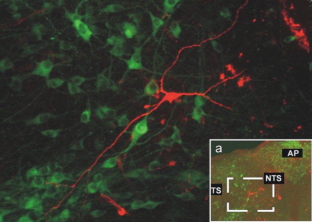
Figure 6.
Postrecording immunohistochemical reconstruction of NTS neuron. The micrograph shows the postrecording immunohistochemical detection in a TH-IR-negative (green, FITC filters) NTS neuron (red, tetramethylrhodamine isothiocyanate filters) from the area depicted in the inset (a). None of the 18 neurons reconstructed were TH-IR positive. TS, Tractus solitarius; AP, area postrema.

