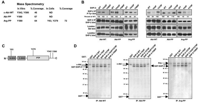
Figure 2. Abl kinases phosphorylate SHP-2 on Y580 and induce phosphorylation of SHP-2 on Y63 and Y279.
(A) GST-SHP-2 was phosphorylated by Abl kinases in vitro, and analyzed by mass spectrometry. For identification of SHP-2 phosphorylation sites in cells, c-Abl and Arg were coexpressed with SHP-2 in 293T cells, and immunoprecipitated SHP-2 was analyzed by mass spectrometry. Coverage is the percent of protein recovered as peptides. ND=not done. (B) Wild-type or mutant forms of SHP-2 and c-Abl or Arg were coexpressed in 293T cells, SHP-2 was immunoprecipitated and probed with phosphotyrosine antibody (top). Percentage phosphorylation is relative to total immunoprecipitated SHP-2 protein obtained from reprobed blots. Results shown are representative of three independent experiments. (C) Structure of SHP-2 and putative Abl kinase phosphorylation sites. (D) Immunoprecipitated Abl kinases were incubated in an in vitro kinase assay with wild-type or mutant forms of GST-SHP-2. Coomassie blue staining showed that SHP-2 proteins were equivalent (data not shown). Results shown are representative of three independent experiments.

