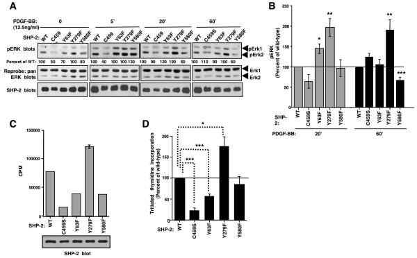
Figure 6. Expression of SHP-2-Y279F increases PDGF-induced sustained ERK phosphorylation and proliferation in fibroblasts.
(A,B) Lysates from serum-starved, PDGF-stimulated NIH3T3 cells, infected with wild-type or mutant SHP-2 retroviruses, were blotted with phospho-ERK1/2 antibody. Changes in ERK1/2 phosphorylation shown are relative to total ERK1/2 levels obtained from reprobed blots. Different exposures are shown for each time point, so that differences between mutants can easily be visualized. Results are expressed as a percentage of wild-type. (B) Results from three independent experiments (A) with mean ± s.e.m. *p≤0.05, **p≤0.02, ***p≤0.005 using t-tests. (C,D) Tritiated thymidine incorporation was assessed in infected NIH3T3 cells labeled with tritiated thymidine for 24 hours. One representative experiment (C) (mean ± s.d.), and a composite of three independent experiments in which tritiated thymidine incorporation of cells expressing SHP-2 mutants compared to cells expressing wild-type SHP-2 is shown (mean ± s.e.m.). Some error bars are too small to be visualized. (D). Aliquots of infected cells were lysed and probed with SHP-2 antibody (C). *p≤0.05, ***p≤0.001 using a one-way ANOVA followed by a Bonferroni post-hoc test.

