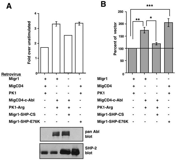
Figure 7. SHP-2 lies downstream of Abl kinases during PDGF-mediated mitogenesis.
c-Abl/Arg double null fibroblasts, infected with vectors (Migr1, MigCD4, PK1), c-Abl and Arg (MigCD4-Abl, PK1-Arg), and/or wild-type or mutant forms of SHP-2 (Migr1-SHP2-C459S, Migr1-SHP-2-E76K), were serum-starved, stimulated with PDGF-BB (12.5ng/ml) for 16-20 hours, pulsed with tritiated thymidine, and harvested. (A) Results from one representative experiment (mean ± s.d). Some error bars are too small to be visualized. Lysates from infected cells were probed with the indicated antibodies. (B) Composite of three independent experiments (mean ± s.e.m). Tritiated thymidine incorporation is expressed as a percentage of vector-transfected cells. *p≤0.05, **p≤0.01, ***p≤0.001 using a one-way ANOVA followed by a Bonferroni post-hoc test.

