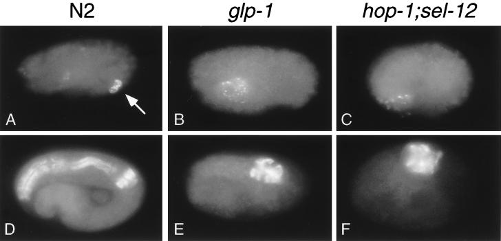
Figure 3.
Phenotypes similar to those observed in a conditional glp-1 mutant are observed in hop-1; sel-12 embryos derived from hop-1; sel-12/+ parent animals. (A–C) Immunofluorescence micrographs of embryos stained with the monoclonal antibody J126 to visualize the intestinal valve cells (17). Wild-type embryos exhibit staining from two intestinal valve cells (arrow in A), whereas neither glp-1(q231ts) (ref. 24 and B) nor hop-1; sel-12 (C) mutant embryos exhibit intestinal valve cell staining. The embryo in C is representative of 25 embryos scored. Either the E or EMS blastomere was laser-killed in each of the embryos shown to eliminate the intestine. The J126 antibody also stains pharyngeal gland cells (17); pharyngeal gland cell staining can be distinguished from valve cell staining based on cell morphology (19). Staining of pharyngeal gland cells is visible in A–C; in A most of this staining is in a different focal plane than that shown. (D–F) Immunofluorescence micrographs of embryos stained with the monoclonal antibody 9.2.1, which recognizes pharyngeal myosin C (20). glp-1 (ref. 23 and E) and hop-1; sel-12 (F) embryos have less pharyngeal tissue than does a wild-type embryo (D). The embryo depicted in F is representative of more than 50 embryos scored. C. elegans embryos are approximately 50 μm long.

