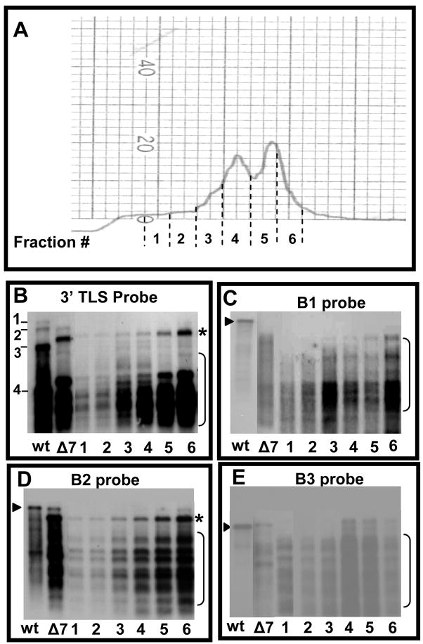
Figure 3.
Fractionation and RNA analysis of polymorphic virions of Δ7aa. (A) Partially purified virions of Δ7aa were subjected density gradient centrifugation as described under Fig. 1D legend. Individual fractions corresponding to areas indicated by dotted lines were collected, concentrated by Centricon-100 columns and RNA was extracted by SDS-phenol/chloroform method. (B–E) Northern blot hybridization. RNA extracted from the each of the six fractions corresponding to Δ7aa slow and fast sedimenting peaks were subjected to multiple Northern blot hybridizations. RNA isolated from unfractionated virions of wt and Δ7aa was used as controls. To maintain uniformity among the RNA sample loaded, each lane contained approximately 100 ng of virion RNA and the blot was hybridized with indicated 32P-labelled riboprobes. The arrow head indicates respective genomic full length RNA whereas the bracketed region indicates truncated RNA species.

