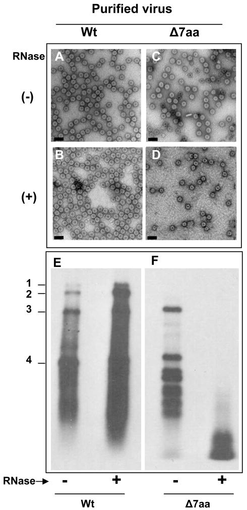
Figure 5.
RNase sensitivity of purified virions. Electron micrographic images of purified virions of wt and Δ7aa following RNase treatment (panels B and D). Untreated virions are shown in panels A and C. Bar = 50 nm. Approximately 50–100μg/ml of purified virions of wt and 7aa were treated with 1μ μg/μl of RNase A for 30 min at 30°C. Following RNase treatment, each preparation was divided into two aliquots. One aliquot was subjected to EM examination while the other was used to extract RNA for Northern blot hybridization (E and F). Preparation of virion samples for EM examination and conditions of Northern blot hybridization are as described under the legends of Fig 1 and 2.

