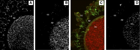
Fig. 3.
Cumulus oocyte complexes (COCs) stained for phosphorylated epidermal growth factor receptor (pEGFR). A: COC from FSH-primed ovary. B: COC from FSH-primed ovary and fixed after 28–30 h of IVM. C: merged image of B showing intact cumulus cells (green, f-actin; red, pEGFR), D: COC from FSH- and human chorionic gonadotropin (hCG)-primed ovary. A–D: pEGFR labeled positive in cumulus cells (cc) and in the oocyte cytoplasm (oc) of all COCs stained. The zona pellucida (zp) was negative for labeling.

