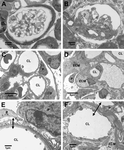
Fig. 7.
Light (A, B) and transmission electron micrographs (TEM; C, D, E, F) of renal cortical tissues from age-matched adult +/+ (A, C, E) and Br/+ (B, D, E) mice. Control (+/+) mice (A) exhibit normal glomerular histoarchitectures, whereas massive increases in extracellular matrix (ECM) are seen in the mutants (B). TEM of normal (C) and mutant (D) tissues illustrate the increase in specific cellularity and ECM in the mutants. Relatively high-magnification TEM of normal (E) and mutant (F) mice show a substantial increase in the blood-urine barrier thickness (double-ended arrows) in the mutants. CL, capillary lumen; P, podoctye. A and B, ×600; C, ×3,000; D, ×2,800; E, ×9,200; F, ×9,100.

