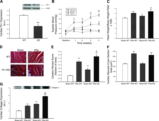
Fig. 1.
Characterization of cardiac fibrosis in Friend leukemia integration-1 (Fli-1)-knockdown (KD) animals and their response to partial nephrectomy (PNx). A: basal Fli-1 expression determined with Western blot (representative blot at top, quantitative data at bottom) in cardiac tissue obtained from n = 5 wild-type (WT) and Fli-1 KD mice. B: conscious systolic blood pressure in WT animals exposed to sham surgery (Sham, n = 6) and PNx (n = 8) as well as KD mice exposed to sham surgery (n = 6) and PNx (n = 9) determined at baseline and 1, 2, 3, and 4 wk. C: heart weight normalized for body weight in these animals at 4 wk. D: representative histology (trichrome stain). E: quantification of fibrosis. F: measurement of myocyte cross-sectional area from the hearts of these mice. G: collagen expression determined with a polyclonal antibody (representative Western blot with dimer at ∼100 kDa, quantified data shown below) in these animals. *P < 0.05, **P < 0.01 vs. Sham-WT; #P < 0.05, ##P < 0.01 vs. PNx-WT.

