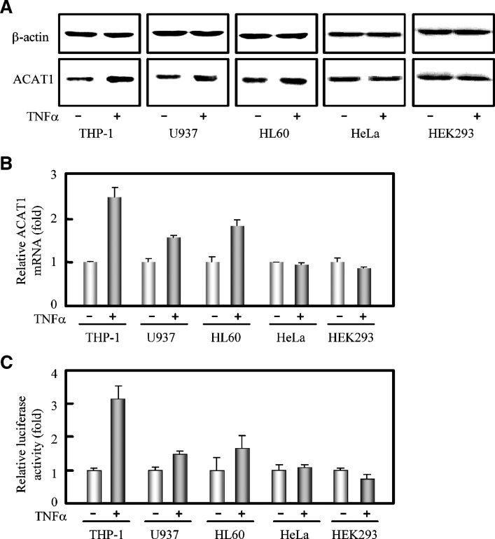
Fig. 4.
Cell type specificity of TNFα in upregulating the expression of human ACAT1 gene. A: Immunoblotting of human ACAT1 protein. Human monocytes (THP-1, U937, and HL60) and nonmonocytic cells (HeLa and HEK293) were directly treated without or with TNFα (5 ng/ml) for 40 h. Immunoblotting was performed as described in “Materials and Methods.” B: RT-qPCR analysis of human ACAT1 mRNA. All the cells were treated the same as in (A). The data were obtained and represented the same as in Fig. 1F. C: Effect of TNFα on the human ACAT1 gene promoter activity. The Luc plasmid pM50 containing the core region (–125/+34) of human ACAT1 gene promoter was cotransfected with pCH110 (for the expression of galactosidase) into human monocytes (THP-1, U937, and HL60) and nonmonocytic cells (HeLa and HEK293) as indicated in “Materials and Methods.” 7 h after transfection, the cells were treated without or with TNFα (5 ng/ml) for another 40 h and harvested for luciferase activity analysis as described in “Materials and Methods.” The luciferase activities were normalized to those of galactosidase in each condition. The data shown as the relative luciferase activities (fold) were calculated from the normalized luciferase activities by setting the average activities of that without TNFα stimulation to 1.0, and represented the means ± SD from triplicate determinations. All of the experiments were repeated three times with similar results.

