Figure 2.
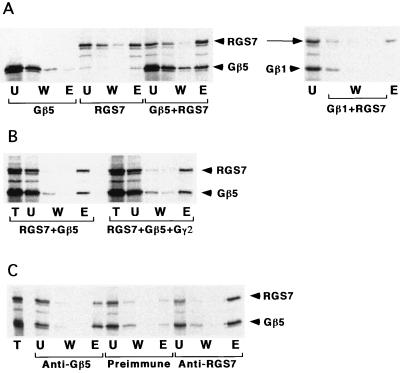
Analysis of RGS7-Gβ complex formation in vitro by cation-exchange chromatography and immunoprecipitation. (A) Chromatography on Sepharose S. The lysates containing [35S]Met-labeled Gβ5, Gβ1, RGS7, or their mixture were incubated batchwise with the chromatography resin. The unbound material was collected, the beads then were washed and eluted by 300 mM of NaCl, and proteins from the fractions were analyzed by SDS/PAGE. T, total lysate loaded; U, unbound material; W, washes; E, the eluate. (B) The Gβ5γ2 complex was obtained by mixing the 35S-labeled Gβ5 and the excess of unlabeled Gγ2 under the same conditions as in Fig. 1. 35S-RGS7-containing lysate then was added to the mixture, and binding to Sepharose S was tested as in A. (C) Immunoprecipitation. The antibodies indicated were adsorbed on protein A-Sepharose, and the lysates were added to the beads. After incubation, the beads were washed and eluted by SDS, and the obtained fractions were processed as in A. T, total mixture added; W, washes; E, eluate.

