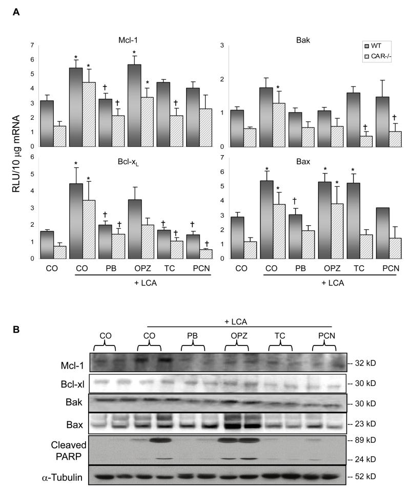
Fig. 3. Apoptosis-related expression in liver.
(A) mRNA expression of anti-apoptotic Mcl-1 and Bcl-xL, pro-apoptotic expression of Bak and Bax as quantified by the bDNA signal amplification assay. N= 4–6 per group, except for WT PCN, N=3. Data are expressed as relative light units (RLU) ± S.E.M. * Indicates p ≤ 0.05 compared to respective CO; † indicates p ≤ 0.05 compared to respective LCA-only. (B) Cytosolic fractions were isolated and analyzed by Western blotting for protein expression of hepatic Mcl-1, Bcl-xL, Bak and Bax, as well as PARP. Two animals per treatment group are shown.

