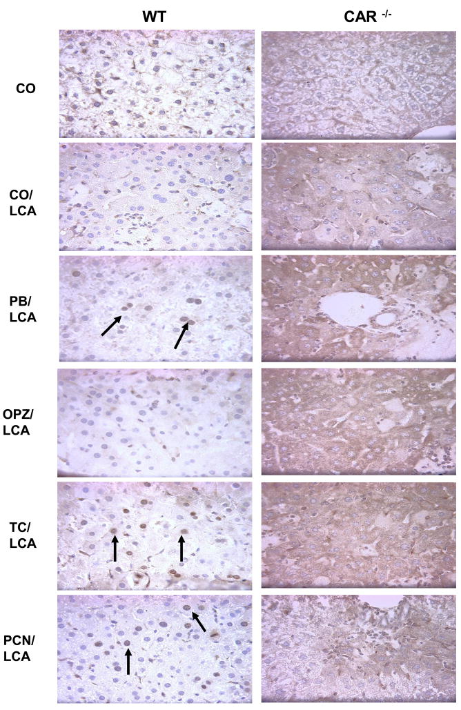
Fig. 5. Immunohistochemical localization of Mcl-1.
The localization pattern of activated Mcl-1 was altered between the groups with hepatocellular damage (cytoplasmic) and those without damage (nuclear). A minimum of two animals per treatment group were evaluated for IHC and pictures are representative of treatment group pathology. 20× magnification.

