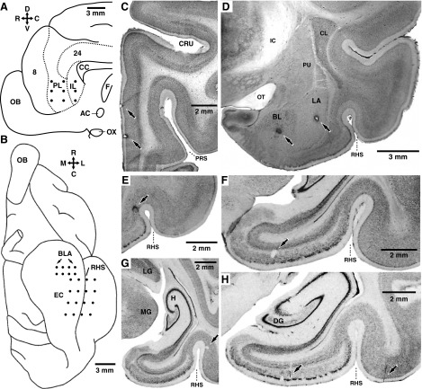
FIG. 1.
Recording method and histological identification of recording sites. A and B: schemes showing a view of the midline (A) or ventral aspect (B) of the cat brain, respectively. •, intended position of microelectrodes in the medial prefrontal (mPFC, A) or basolateral amygdala (BLA), entorhinal (EC), and perirhinal cortex (B). C–H: histological verification of recording sites. Coronal brain sections showing the location of electrolytic lesions (↑) performed at the end of the experiments to mark the position of the microelectrode tips in the mPFC (C), BLA (D), perirhinal cortex (E, G, and H), and entorhinal cortex, (F and H). The orientation of A and B is indicated by crosses where R, C, D, V, M, and L stand for rostral, caudal, dorsal, ventral, medial, and lateral, respectively. AC, anterior commissure; CC, corpus callosum; CL, claustrum; CRU, cruciate sulcus, BL, basolateral nucleus of the amygdala; DG, dentate gyrus; F, fornix; H, hippocampus; IC, internal capsule; IL, infralimbic area; LA, lateral nucleus of the amygdala; LG, lateral geniculate nucleus; MG, medial geniculate nucleus; OB, olfactory bulb; OT, optic tract; OX, optic chiasm; PL, prelimbic area; PRS, presylvian sulcus; PU, putamen; RHS, rhinal sulcus.

