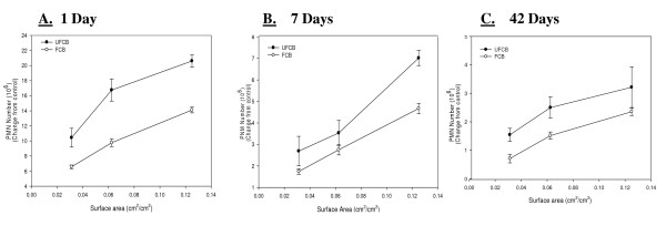
Figure 2.
Comparison of inflammation elicited in animals receiving UFCB and FCB normalized to surface area of particles administered per surface area of alveolar epithelium. A comparison of inflammation elicited in animals receiving doses (0.0313, 0.0625 and 0.125 cm2/cm2) of UFCB and FCB normalized to surface area of particles administered per surface area of alveolar epithelium at 1 day (Panel A), 7 days (Panel B), and 42 days (Panel C) post-exposure. Particles were suspended in BALF. Alveolar epithelial surface area for the rat was taken from Stone et al. [14]. Rats were exposed to various doses of UFCB and FCB by intratracheal instillation. Animals were euthanized at 1 day, 7 days, and 42 days post-exposure and bronchoalveolar lavage was performed. Inflammation was assessed by BAL PMN counts. Values are increased PMN number above the BALF control and are given as means ± SEM of 8 rats. Control PMN values were 1.45 ± 0.22 × 10⁶, 1.09 ± 0.14 × 10⁶, and 1.01 ± 0.14 × 10⁶ cells/rat for 1, 7 and 42 days respectively. Linear regression analysis with a 95% confidence interval reveals that when dose is normalized to surface area of particles administered, dose responses curves assessing inflammation caused by UFCB and FCB exposure are not significantly different. On a dose normalized to surface area UFCB elicits at most a 2 fold increase in inflammation when compared to FCB at all post-exposure time points.

